
by Ellen Stodola
Editorial Co-Director
Corneal tissue procedures are one of the approaches available to help treat corneal conditions, like keratoconus, and offer some patients an option other than transplantation. Several experts discussed these procedures, how they fit in with other options, and how they’re being utilized today.
For keratoconus, there were no good treatments for a long time, said Jack Parker, MD, PhD, but in the past 10 years, there have been some new developments. For a long time, the only thing we had for keratoconus patients was to offer them a full corneal transplant, and PK is not necessarily a good option for people with keratoconus because these are young people, he said, adding that it’s an invasive operation with lifetime risk and obligation. There’s been a longstanding desire for something better. The initial effort to fix that was DALK, but DALK is still invasive and aggressive, he said.

Source: Soosan Jacob, MD
There’s also crosslinking, Dr. Parker said, which will stop the progression of disease, but it doesn’t make vision better. CAIRS (corneal allogenic intrastromal ring segments) and CTAK (corneal tissue addition keratoplasty) are exciting because these are surgical options that can make patients’ vision better, but they don’t come with all the potential risks.
CTAK and CAIRS represent a fundamentally different philosophy compared to traditional approaches, said Aylin Kiliç, MD. Conventional treatments like synthetic intracorneal ring segments (e.g., Ferrara Ring [Ferrara Ophthalmics] or Intacs [Addition Technology]), corneal crosslinking, or penetrating keratoplasty halt progression, mechanically reshape the cornea with foreign material, or replace it entirely.
“CTAK and CAIRS, by contrast, use donor human stromal tissue to reinforce and reshape the cornea from within, essentially augmenting the patient’s own cornea rather than replacing or artificially propping it,” she said. “This biologic approach preserves the native tissue architecture, maintains a more natural biomechanical profile, and avoids the immunological and mechanical complications associated with synthetic materials. They occupy a critical middle ground between crosslinking and transplantation that was previously underserved.”
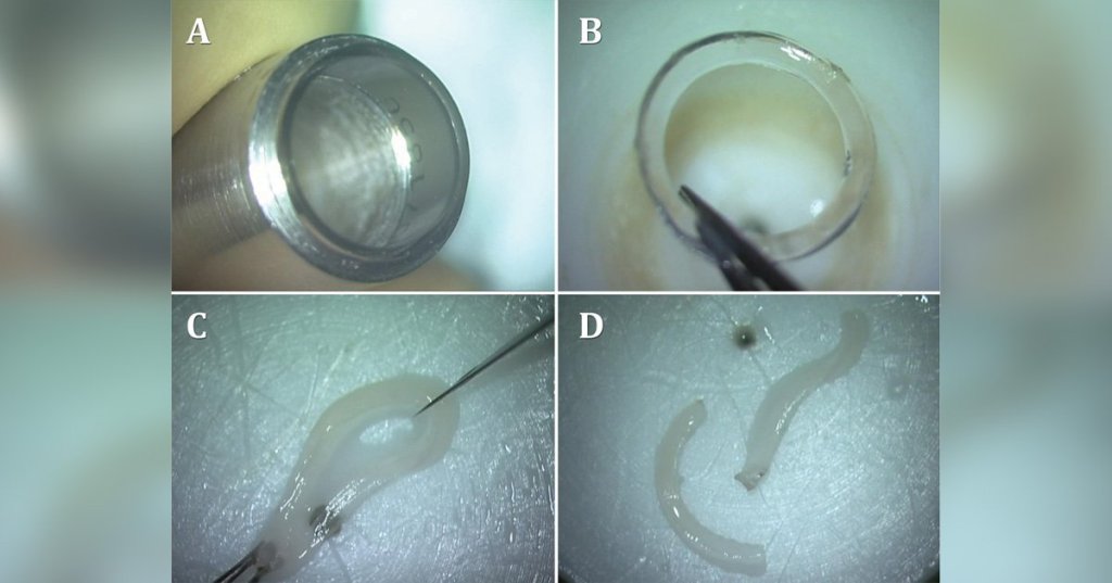
Soosan Jacob, MD, first clarified the terminology and history of these procedures, noting that she started using CAIRS around 2015. She was doing uniform thickness CAIRS before transitioning to customized and custom-shaped CAIRS in 2017. You make mid-peripheral femtosecond laser channels and implant tissue segments into those channels. The whole purpose is to change the shape of the cornea.
She also noted the various types of CAIRS now available, including ECO-CAIRS, decellularized CAIRS, and CTAK. CTAK, which has become available within the U.S., started with clinical trials in 2015 as a disk that was placed within a pocket in the cornea and not as mid-peripheral segments in channels. A 2023 paper published in the Journal of Cataract & Refractive Surgery1 described CTAK as a new procedure, however, many physicians performing CAIRS label CTAK as a branded subtype of CAIRS since variations such as femtosecond cutting, customization, and decellularization were already being performed for many years with CAIRS. In addition, only conventional CAIRS currently uses custom shaping to enhance personalization for every eye, she said. Dr. Jacob noted that she uses CAIRS frequently in her practice.
Adoption is growing, Dr. Kiliç said, but is still relatively limited to specialized corneal centres and early-adopter surgeons globally.
Dr. Parker said he is also utilizing CAIRS frequently in his practice. It’s not uncommon for him to do six or seven a week. However, he agreed that these options haven’t quite hit the mainstream in the U.S.
Patients with central scarring are usually not very impressed by the results with CAIRS or CTAK, he said, because even if the shape of the cornea is better, there are still reasons why they can’t see through it in those with extremely advanced disease.
Performing the procedure
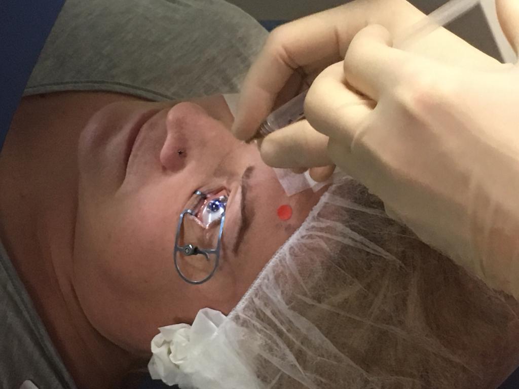
There are a couple of ways to do these procedures, Dr. Parker said. The first is to do a femtosecond laser-assisted technique, which is generally straightforward, and many surgeons are familiar with the operation. Once the channels where these segments are placed are created, it’s relatively straightforward, he said. That’s a convenient way for many surgeons to start, especially if they already have the laser.
The alternative is a manual strategy for creating the channel. Manual dissection has a steeper learning curve and presents a bit more of a challenge for the starting surgeon, Dr. Parker said. However, it doesn’t use the laser, so for surgeons who don’t have access to the laser or who don’t want to use the laser, the manual technique is a way to get started.
Dr. Jacob mentioned a comparison to synthetic segments, however, she noted that these can be prone to melt, so you have to be careful to have a lot of stroma above a synthetic segment. Because of this, it’s placed very posterior. You can’t treat patients below a certain thickness. In CAIRS, you can decrease the optical zone, implant thicker segments, and implant more superficially, Dr. Jacob added, which allows you to treat more advanced cases.
While both allogenic and synthetic segments allow for customization, Dr. Jacob said synthetic options may only be customized with linear gradation and not as true customization because “customized” synthetic segments are manufactured in stock sizes. With CAIRS, she has described custom shaping since 2017, and you can individually shape it for each eye of a particular patient. Keratoconus is never the same in any two patients or two eyes, so there’s no way that you can treat it without customizing, and custom shaping is simple with her nomogram, she said.
Dr. Jacob noted she uses the double-bladed trephine (Jacob CAIRS trephine, Madhu Instruments) that is available in various sizes to cut the tissue, and this provides results that are just as accurate as using a laser because of the precise computer numerical control nature of the manufacturing technology. The added advantage is that you can get the full thickness of the tissue, unlike with the femtosecond laser, which allows only partial thickness cuts, she said. This allows potentially greater flattening with the special double-bladed trephines while still allowing for the tissue to be split and used if required. “Another big advantage of manual shaping is that you can customize it exquisitely,” she said. With the femtosecond laser, you can customize, but you would need a lot of calculations, and even then, there are certain limitations.
There are several differences to using an allogenic inlay to a synthetic option, Dr. Parker said. He finds that one advantage of an allogenic is it’s not a foreign material that the body is trying to spit out. “When you have something like a piece of plastic, the body may try to reject that,” he said, and it can extrude out through the front surface of the eye over time. When that occurs, it could be a disaster with corneal melting and potential infection.
With allogenic inlays, the body doesn’t have that same natural tendency to reject them. As a result, he thinks that’s safer to put inside of the eye. “Furthermore, you can use bigger segments, which have a greater topographic effect, and you can place them more anterior in the cornea, which also has a more topographic effect,” he said. Finally, you can use them in thinner corneas. With CAIRS and CTAK, you can place big segments higher in the cornea, and in patients with much thinner corneas, you get much more of an effect.
The distinction between allogenic inlays and synthetic inlays is both biological and biomechanical, Dr. Kiliç said. Synthetic inlays—such as the Keraring (Mediphacos), Intacs, or Ferrara Ring—are made from materials like PMMA or hydrogel polymers. They function by shortening the arc, creating a mechanical spacer effect within the stroma, redistributing tissue tension, and altering corneal curvature, she said. “However, they remain a foreign body, which carries risks of interface haze, extrusion, infection, and long-term material degradation. They do not integrate with the host tissue.”
She noted that allogenic stromal inlays, by contrast, are composed of human collagen—they are biologically compatible with the host stroma and, over time, integrate into the native extracellular matrix. “This integration means the reshaping effect is maintained by tissue, there is no foreign body inflammatory response, and the biomechanical behavior is far more physiologically appropriate. The cornea essentially incorporates the donor tissue as part of its own architecture. This integration is a major theoretical and practical advantage.”
Best candidates and success rates
The ideal candidates, Dr. Kiliç said, are patients with progressive keratoconus or post-refractive ectasia who still have enough clarity to avoid transplantation. Specifically, she noted patients with moderate to advanced keratoconus, corrected distance visual acuity that is still compromised despite spectacles or contact lenses, and a desire to defer a penetrating or lamellar keratoplasty are strong candidates. This can also be combined with crosslinking for added stability.
Early and mid-term data are encouraging, Dr. Kiliç said, noting that published studies on CAIRS report meaningful improvements in uncorrected and corrected visual acuity, significant reductions in keratometry readings (often 3–6 D of flattening), and improvements in higher order aberrations. Patient satisfaction tends to be high as well, she said. In well-selected patients, these procedures appear capable of deferring or potentially avoiding keratoplasty in a meaningful proportion of cases.
“That said, long-term data beyond 5–7 years remains limited, and we don’t yet have robust randomized controlled trials comparing these techniques head-to-head with DALK or PKP,” Dr. Kiliç said. “Success is also highly dependent on patient selection—in very advanced ectasia with central scarring, these additive approaches are unlikely to suffice.”
According to Dr. Kiliç, short-to-medium-term results are genuinely promising—improvements in visual acuity, topographic flattening, and reduced dependence on rigid contact lenses are consistently reported. Complications reported in the literature include interface haze (generally mild and transient), partial lenticule displacement in early cases, and suboptimal centration. Unlike synthetic rings, there are limited reports of extrusion of allogenic material to date, which is a meaningful safety advantage.
The best candidates are patients with keratoconus or ectasia at a stage where some stromal reinforcement and reshaping is needed but the cornea still has adequate transparency and thickness, she said. Contraindications include corneas with significant central scarring (which would benefit more from a transplant), active ocular surface disease, significant dry eye, or corneas below the minimum safe residual stromal thickness for tunnel creation.
The data for the predecessor operation, Intacs, suggested that two-thirds of patients who had these segments implanted got two lines of vision or better on the eye chart, Dr. Parker said, adding that CAIRS is a little better than that. With carefully selected patients, we often are very impressed with the results. It’s rare for patients to have CAIRS and end up needing a transplant after that because of disappointing visual outcomes. That said, if you’re operating on someone who is hand motion, and they’re not going to be satisfied with 20/100 vision, you might find yourself doing more interventions, Dr. Parker said.
Dr. Jacob generally finds good success with these procedures, although she did note that this is therapeutic refractive surgery, so it can sometimes be hard to define “success” with a specific number or outcome. It’s not necessarily like SMILE or LASIK, she said, where you’re aiming to get the patient free of glasses. With this, you’re aiming for improving quality of vision and not glasses-free vision.
CAIRS, she said, can not only help with improving the topography and vision but may also possibly play a role in improving biomechanics.2 To an extent, it can also help with the stabilization of the disease because of the redistribution of the biomechanical stress forces, so the cornea becomes more regular. Just the simple redistribution can also make the corneal biomechanics stronger, she said. So, the chances of needing a corneal transplant may decrease. Another benefit of CAIRS is you can adjust it or reverse if the patient is unhappy.
In terms of concerns and contraindications, Dr. Jacob said it’s applicable for a large majority of the patients, but there may be some extreme cases, like very ectatic corneas, where it might not be a good option. In extremely ectatic corneas, which are too thin to crosslink, Dr. Jacob might still consider other procedures such as DALK. If there is a central scar over the visual axis, Dr. Jacob might also choose DALK instead of CAIRS. She added that in patients with autoimmune disorders, there may be some concern about implanting tissue in the cornea.
Another point is that when you implant the tissue, you should not overstuff, she said. When starting out with this procedure, some may be inclined to try to make narrow tunnels and stuff a lot of tissue into them. But if you keep stretching, at a point, it will give way. Excessive stuffing and extremely narrow tunnels should be avoided, she said, and it’s important that the segments fit comfortably within the tunnels.
Looking forward
There have been many publications from authors around the world on this, Dr. Jacob said, adding that there have been positive results in uncorrected and best corrected VA with CAIRS. She noted a study by Gerd Auffarth, MD, PhD, that is a meta-analysis of all studies that shows results have been good.3
Dr. Jacob noted that CAIRS is being used in more than 40 countries worldwide. “It’s surgery with a low risk and high reward.” This can be used in young patients, since there are risks with keratoplasties and other procedures. “I think that’s what has made CAIRS so popular,” she said.
Dr. Jacob advised surgeons who are interested in learning this technique to take a course or watch videos to learn more. The more important learning curve is in the planning, she said, adding that she has her own proprietary nomogram which she uses for custom shaping, and there are other nomograms available.
At present, these procedures are gaining increasing interest worldwide, Dr. Kiliç said, but they are not yet universally adopted. “One of the main limiting factors is the requirement for a femtosecond laser, particularly for precise channel creation,” she said. “The lack of access to this technology remains one of the most important barriers to broader utilization.”
Although commercially available products such as KeraNatural (VisionGift) have simplified the process, Dr. Kiliç said it is important to note that surgeons also have the option to prepare allogenic tissue intraoperatively. But this approach requires additional surgical skill, familiarity with tissue handling, and a certain level of adaptation within the operating room, often supported by dedicated training programs.
“For surgeons who already have access to femtosecond laser platforms, the learning curve is relatively manageable,” she said. Like Dr. Jacob, Dr. Kiliç pointed out the importance of surgical planning, adding that the Istanbul nomogram is now widely recognized and has become one of the most practical and user-friendly nomograms in the field. Industry support also plays an important role in accelerating the learning process, she said.
“Overall, while there are certain technological and educational barriers, the combination of standardized products, simplified nomograms, and increasing training opportunities is steadily improving accessibility and adoption,” Dr. Kiliç said.
One aspect that she is excited about is the potential to significantly reduce the need for keratoplasty in keratoconus patients. Inlay- based approaches, such as allogenic corneal implants, represent a less invasive, adjustable, and tissue-preserving alternative compared to procedures like DALK, she said.
“We have already made remarkable progress, but I think that this is only the beginning. With the accumulation of larger datasets and more refined analysis, we will be able to achieve more precise and predictable outcomes,” Dr. Kiliç said. “Keratoconus is a highly heterogeneous disease—each cornea is unique, and each patient may respond differently to the same treatment. This variability is one of the main challenges we still face today. However, as we improve our ability to predict these responses, I think these techniques could evolve toward a refractive, highly customized approach, ultimately allowing us to provide better visual outcomes for a broader group of patients.”
From Cornea
Summer 2026

Leave a Reply